Machine Learning-Aided Non-Invasive Imaging for Rapid Liver Fat Analysis
Share
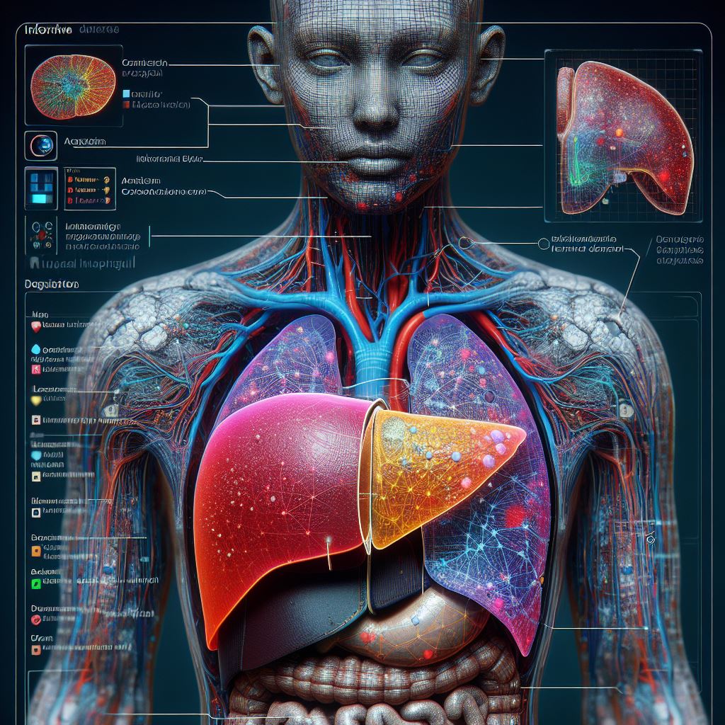
Steatotic Liver Disease (SLD), previously recognized as non-alcoholic fatty liver disease, encompasses various conditions resulting from an abnormal accumulation of fat in the liver due to disrupted lipid metabolism. Affecting approximately 25% of the global population, it stands as the most prevalent liver disorder. Often termed "silent liver disease," SLD advances without noticeable symptoms and poses the risk of progressing to severe conditions like cirrhosis and liver cancer.
Traditionally, liver biopsy, an invasive procedure involving the extraction of liver tissue samples, has been the standard method for SLD testing. To streamline detection, a research team led by Professor Kohei Soga from Tokyo University of Science (TUS) previously introduced near-infrared hyperspectral imaging (NIR-HSI) as a non-invasive means to visualize total lipid content in the liver. NIR light, with longer wavelengths than ultraviolet and visible light, reveals absorption characteristics of various organic substances, facilitating the identification of fat distribution in the liver.
In a recent study published in Scientific Reports on November 23, 2023, the research team, including Prof. Kohei Soga, Associate Professor Masakazu Umezawa, and Associate Professor Masao Kamimura from TUS, and Professor Naoko Ohtani from Osaka Metropolitan University, has enhanced this method by incorporating a machine learning model to discern the type of lipids present in the liver at a pixel-by-pixel level. This framework distinguishes lipids based on hydrocarbon chain length (HCL) and degree of saturation (DS) of fatty acids, aiding in estimating the risk of SLD progression, steatohepatitis (NASH), and SLD/NASH-associated liver cancer.
Dr. Umezawa explains, "In addition to qualitative information, such as the total lipid content, we can now also visualize qualitative information, such as the characteristics of the distribution of fatty acids contained in lipids, mainly triglycerides."
The researchers addressed challenges in identifying lipids based on molecular composition using NIR-HSI by employing a support vector regression machine learning model. Trained to recognize the composition of 16 fatty acids using data from gas chromatography analysis of mouse liver samples, the model enabled the interpretation of spectral information regarding fat distribution within the liver.
Double bonds or the degree of saturation of fatty acids (DS) and fatty acid chain length (HCL) were calculated, and the total lipid content was represented as a color map, providing a distinctive visual representation of fat distribution in the liver. This approach simplifies the diagnosis of fatty liver conditions, offering a potential alternative to invasive liver biopsy procedures and transforming liver care.
The novel framework also holds promise in pharmacological research, metabolic imaging for studying metabolic disorders, and identifying personalized nutritional strategies. By offering a rapid and label-free technique for identifying fatty liver, this method has the potential to revolutionize healthcare and related research.


